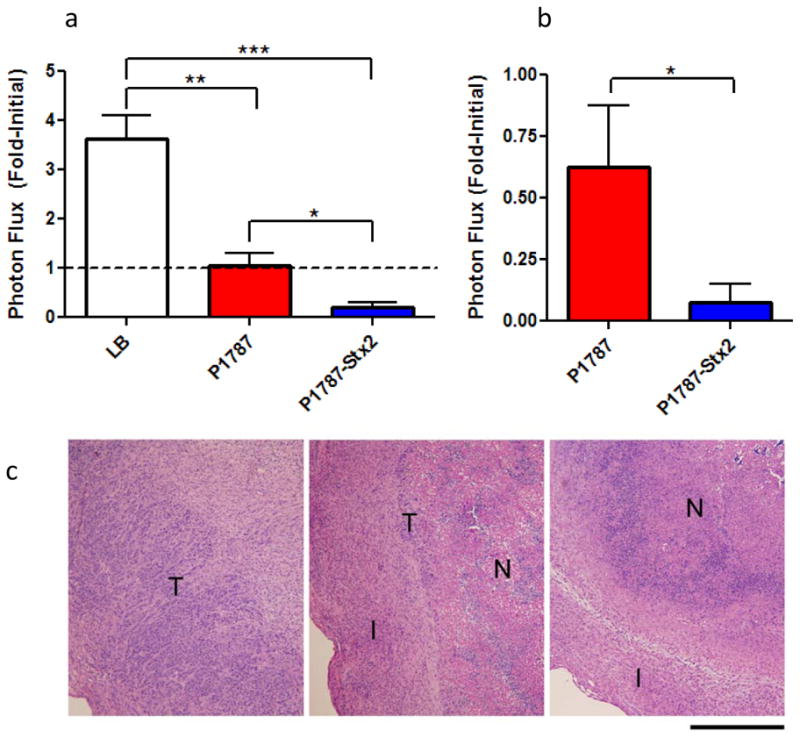
Figure 6. Enhanced anti-tumor response with P1787-Stx2 in vivo.

(a) Viable cell mass of HeLaCMV-FLuc tumors from mice treated with LB (n = 14), or high-dose SB300A1 transformed with P1787 (n= 12) or P1787-Stx2 (n = 9) at five days post treatment. Results are combined from two independent experiments and presented as fold-initial photon flux. Dotted line demarks lack of fold-change in tumor bioluminescence. Error bars indicate standard error of the mean ***p<0.0002,**p<0.0003, *p<0.007). (b) Fold-initialized photon flux of HeLaCMV-FLuc tumors from mice treated with low-dose SB300A1 transformed with P1787 (n= 7) or P1787-Stx2 (n = 7) at 14 days post treatment. Error bars indicate standard error of the mean.*p <0.04. (c) H&E staining of HeLaCMV-FLuc tumors from mice treated with LB (left), or high-dose SB300A1 transformed with P1787 (middle) or SB300A1 transformed with P1787-Stx2 (right) after five days. Regions of tumor are denoted as tumor (T), fibroinflammatory reaction (I), and necrotic zone (N). Scale bar, 500 μm.
