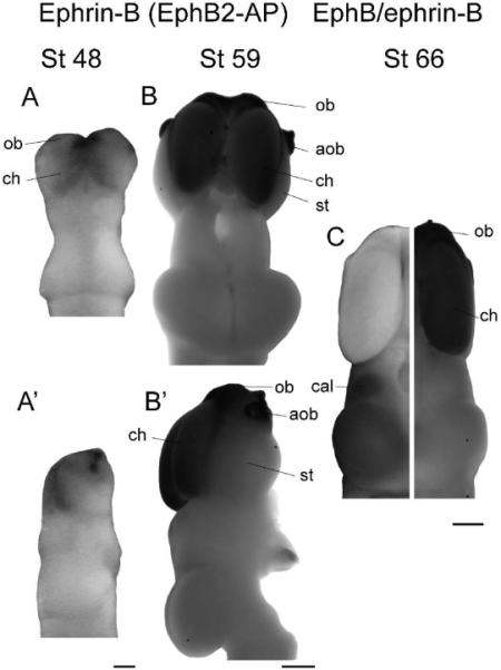
Figure 5. Ephrin-B expression.

(A,B) Dorsal views of stage 48 (A) and 59 (B) brains stained for ephrin-B expression. (A',B') Side views of the same brains. Ephrin-Bs are expressed very strongly in the cerebral hemispheres and olfactory regions at all stages. In older animals specifically, strong expression can be seen in the olfactory bulb, accessory olfactory bulb, cerebral hemispheres, and at the midline of the optic tectum, although staining is undetectable in the striatum and the tectal lobes. (C) Composite image showing EphB (left) and ephrin-B (right) expression in the stage 66 brain. The overall expression patterns of EphBs and ephrin-Bs are complementary; the telencephalon stains for ephrin-Bs, while the diencephalon and optic tectum show staining only for EphBs. Scale bars: A,A'=100 μm, B-C=500 μm.
