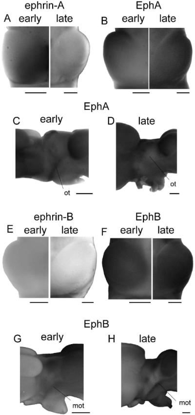
Figure 6. Expression of Ephs and ephrins in adult Xenopus.

(A,B) A composite image of tectal lobes from early (stage 66, left) and late (right) postmetamorphic animals stained for ephrin-A (A) and EphA (B) expression. The very striking gradients present in early postmetamorphic animals are faint if not absent in the late postmetamorphic brains stained either for EphAs or ephrin-As. (C,D) Side view of EphA expression in an early (stage 66; C) and late postmetamorphic brain (D). EphAs appear to be expressed in the optic tract at stage 66, but do not persist in the late postmetamorphic animal. (E,F) Tectal lobes from early (stage 66, left) and late (right) postmetamorphic animals stained for ephrin-B (E) and EphB (F) expression. The very low expression of ephrin-Bs and very high expression of EphBs appear to continue unchanged into adulthood. (G, H) The side view of an early (stage 66; G) and late (H) postmetamorphic brain stained for expression of EphBs. EphBs are strongly expressed in the marginal zone of the optic tract in both froglet and fully mature animal, but ephrin-Bs were not detected in the optic tract at any time point. Scale bars = 500 μm.
