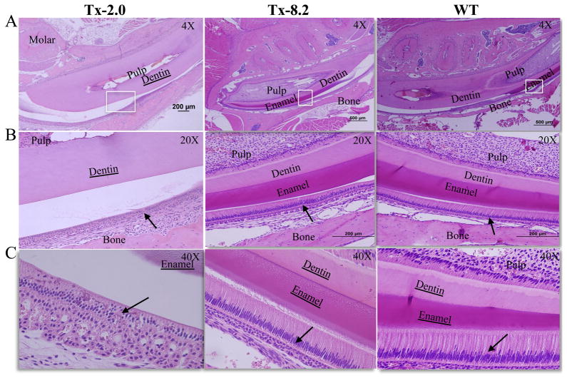
Figure 3.
Histological analysis of enamel development in molars and incisors of Alpl−/− mice with enzyme replacement treatment. H&E staining of mandibles from (A, B and C) 44 dpn Alpl−/−mice given Tx 2.0, Tx-8.2 and untreated WT mice. Enamel seemed to be already formed in the Tx-2.0 group, and the TX-8.2 group shows complete prevention of enamel defects at 44 days of age. At this stage the mature enamel of the incisor in Tx-8.2 group was comparable to that in WT mice. Loss of organization of ameloblasts and disorganized enamel matrix ECM production in the Tx-2.0 group was drastically improved in the Tx-8.2 group, as the enamel organ was rescued to resemble what is seen in WT mice (B and C, arrows). White boxes indicate the corresponding region from A to B and C.

