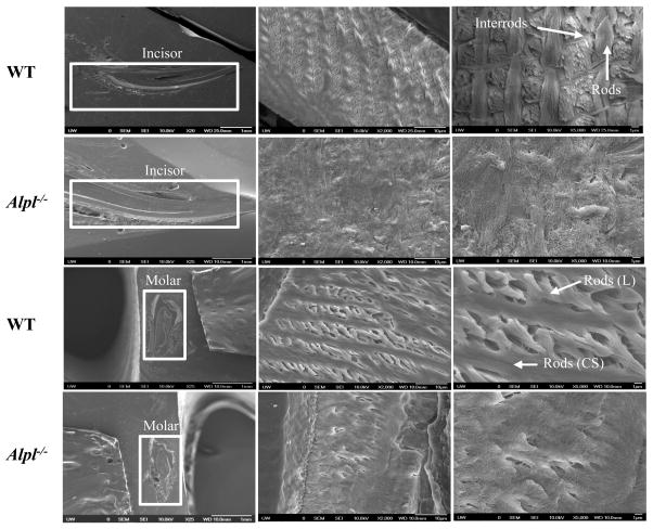
Figure 5.
Scanning electron microscopy (SEM) analysis of incisors (top) and molars (bottom) of WT and Alpl−/− mice at 20 dpn. The SEM images showed well-decussated enamel rods and inter-rod in the molar crowns and crown analogs of incisors of WT mice. Note that there is lack of rod-interrod organization in the Alpl−/− mice. The images were taken in the erupted part of the tooth.

