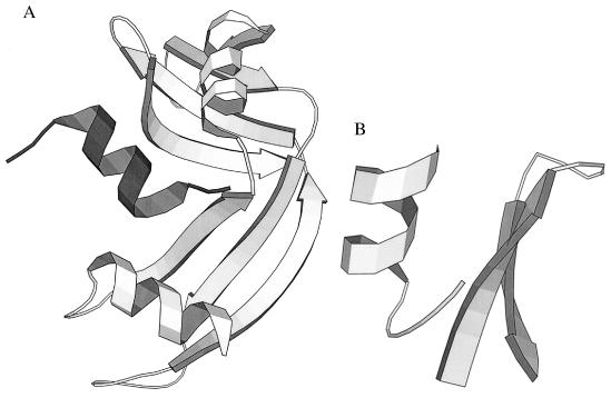
Figure 2.
(A) X-ray structure of ribonuclease S (2RNS) (25). S-peptide (residues 1–20, dark shading), a helix, has been cleaved from S-protein (residues 21–124, light shading), the remainder of the molecule. (Residues 16–23, which are disordered in the crystal structure, are not shown.) (B) X-ray structure of two fragments from intact BPTI (1BPT) (26). The β-hairpin fragment, Pβ, comprises residues 20–33, and the helical fragment, Pα, comprises residues 43–58. Apart from these two segments, the rest of the molecule is not displayed.

