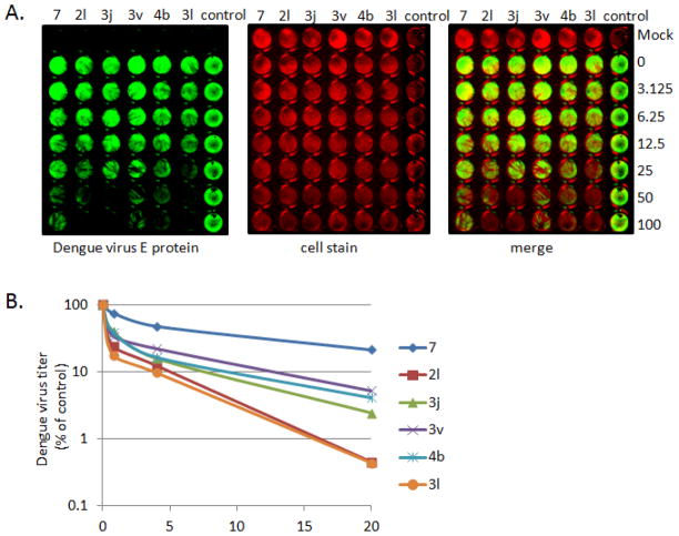
Figure 5.
Side-by-side comparison of anti-DENV activity in in-cell western assay and yield reduction assay. (A) Huh 7.5 cells were infected and treated with indicated compounds, as described in “Experimental Section”. Green color represents the detection of DENV E protein, red for cell stain. (B) Standard yield reduction assay was performed using indicated concentration of compound. DENV titers were determined and expressed as % of control. Values plotted represent the average of three experimental repeats.

