Figure 1.
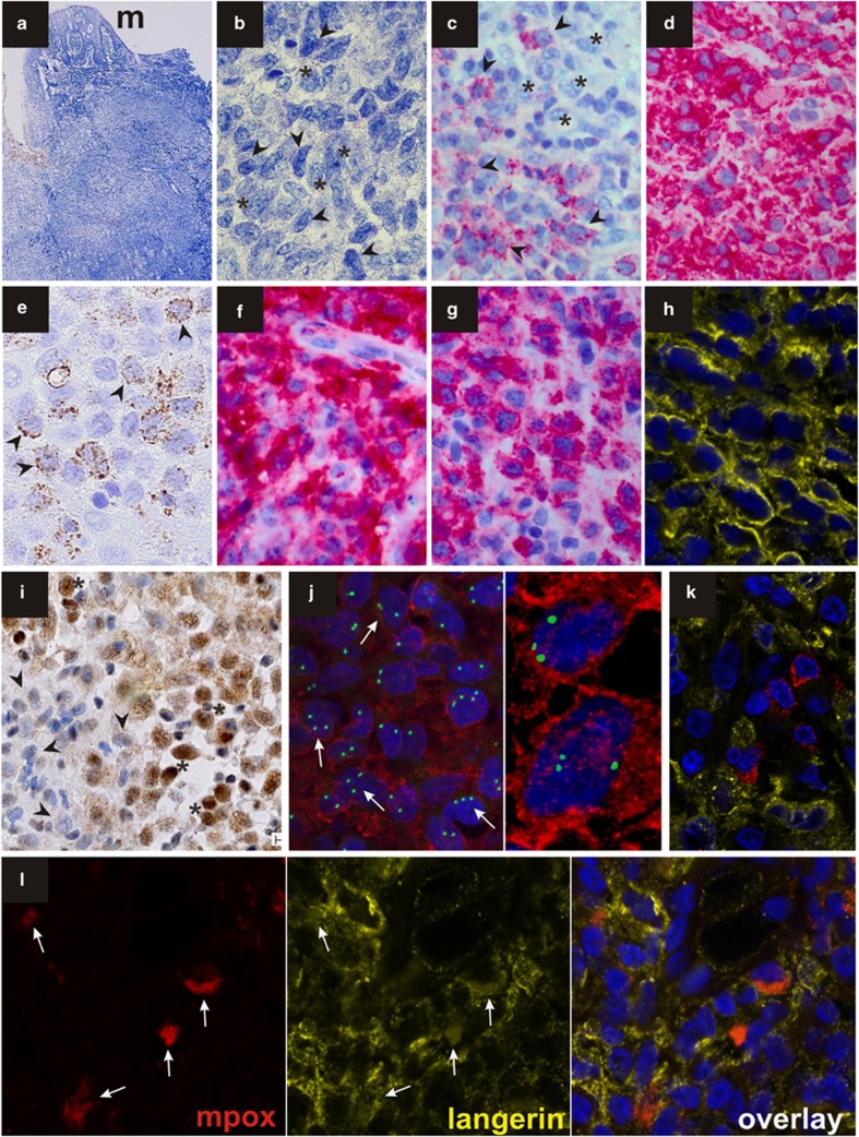
(a) Histologic aspect of the Giemsa-stained gingival biopsy characterized by a broad-based mass located beneath the oral surface mucosa (m). (b) Large LCHCs with grooved nuclei and pale cytoplasm (asterisks) as well as smaller blasts with oval or delicately folded nuclei and indistinct nucleoli (arrowheads) were observed (Giemsa). The blasts (arrowheads) expressed myeloperoxidase (c), CD 33 (d) and showed a finely granular CD68 staining (e). While LCHCs were negative for myeloperoxidase (c, asterisks) they were labelled by CD1a (f), langerin (g, h, confocal image) and Id2 (i) antibodies. By FISH analysis using a DNA probe for trisomy 8 (green) and immunstaining for langerin (red), a trisomy 8 (arrows) was clearly detected in LCHCs (j). CLSM imaging of double stains for myeloperoxidase (red) and langerin (yellow) showed the presence of scattered myeloperoxidase-positive blasts within the sheets of langerin-positive LCHCs (k). There was evidence of a rare coexpressions of both antigens in identical cells, which were stained both in red and in yellow (l; arrows).

