Figure 3.
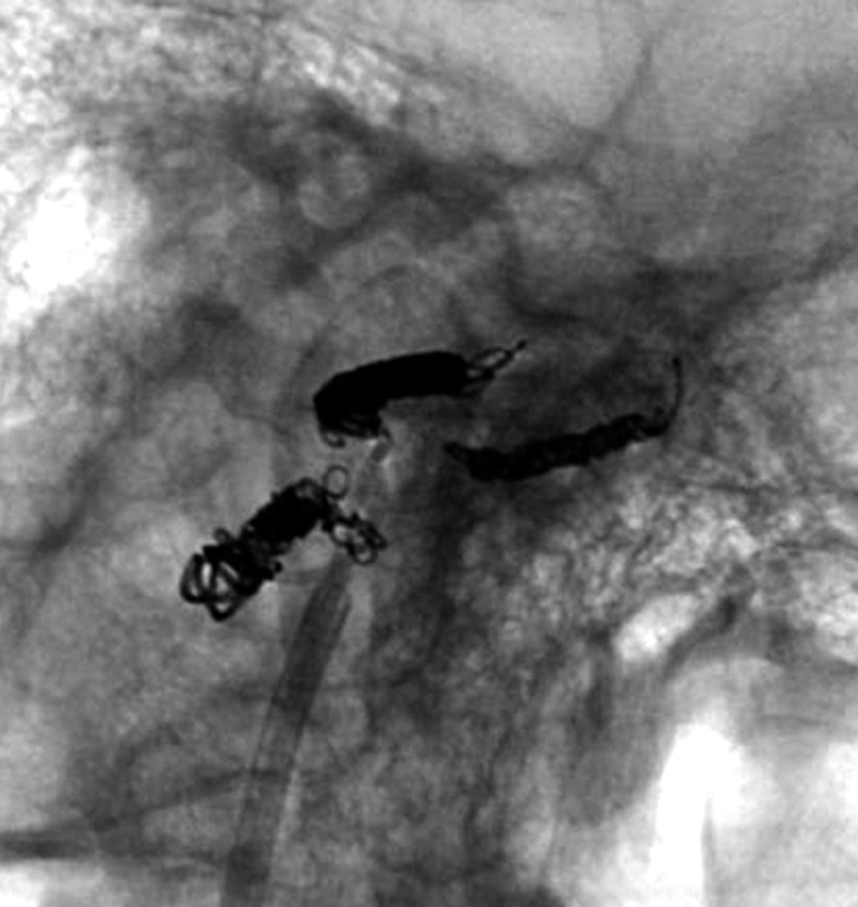
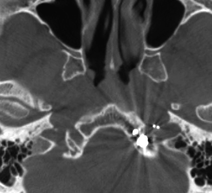
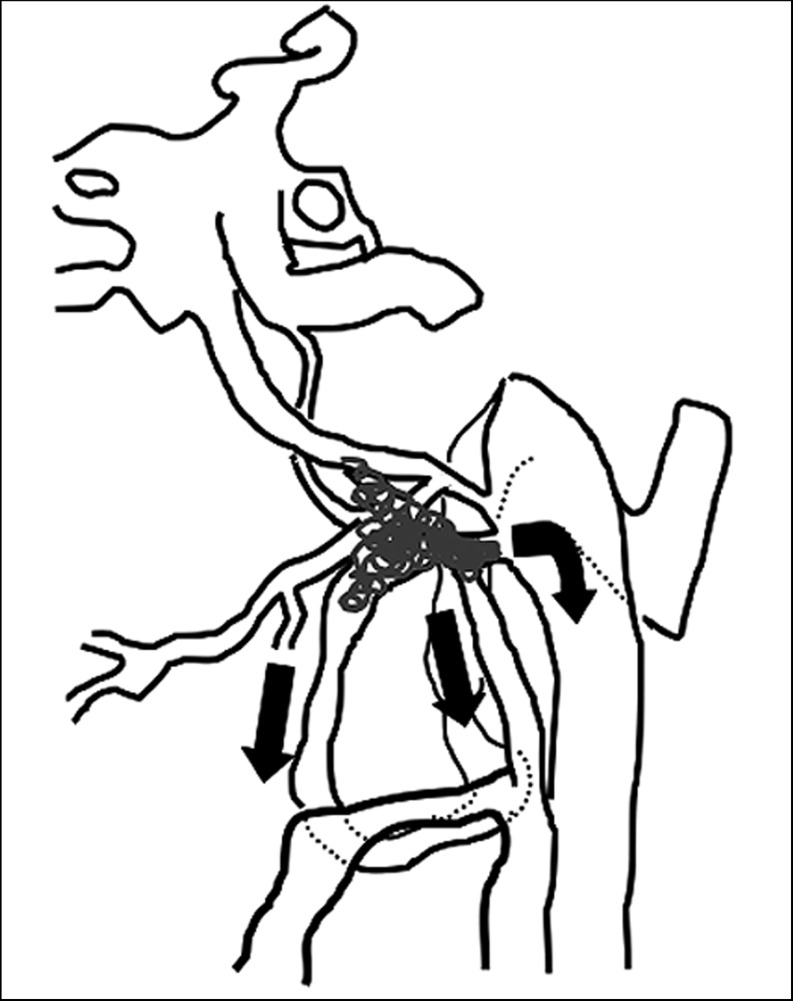
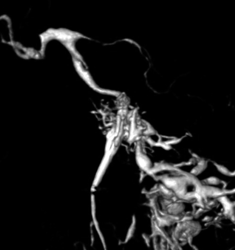
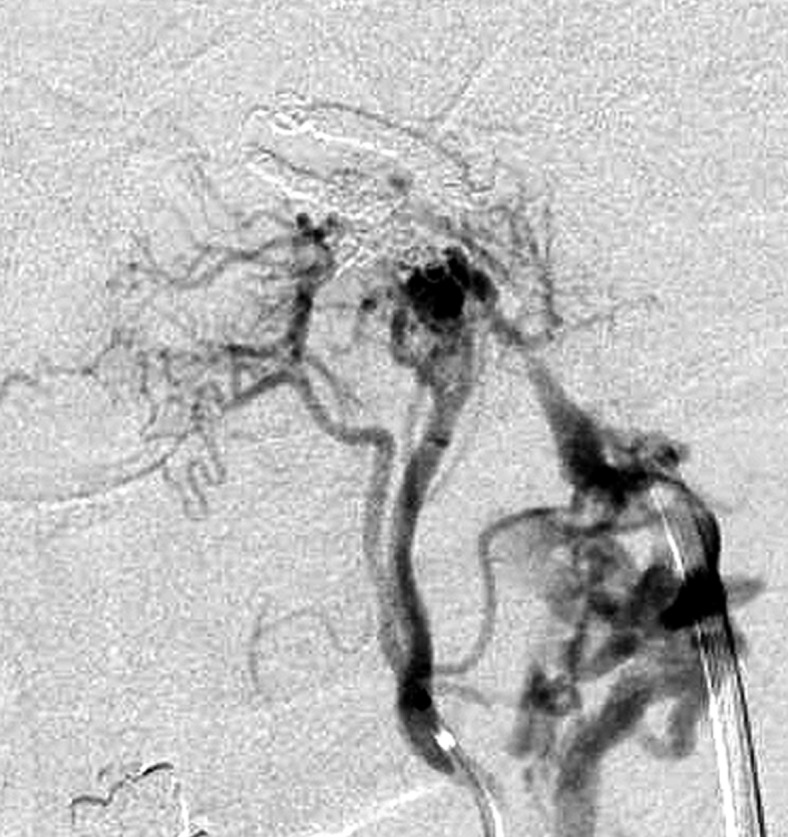
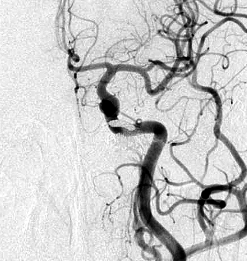
Case 4. Selective 3-D angiogram of left ascending pharyngeal artery (APA) showing the DAVF at the ACC (A). Selective APA angiogram after embolization of the draining veins (inferior petrosal sinus and Trolard’s inferior petro-occipital vein) showing the remaining drainage into the vertebral plexus through the lateral condylar vein (LCV) (B). Coils were added into the LCV (C) and total occlusion of the DAVF was achieved (D). Postoperative Dyna-CT shows the coils placed to occlude the IPS at the superolateral side of the hypoglossal canal (E). Scheme of the shunt point and embolized portion (F).
A.
B.
C.
D.
