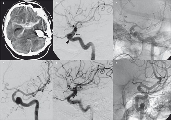
Figure 1.
Case 1. A) Acute CT scan showing diffuse subarachnoid hemorrhage. B) Lateral view of the left carotid artery showing the 8 mm intracavernous aneurysm (arrowhead) and the 3 mm BBL aneurysm of the supraclinoid siphon (black arrow). C) The 7 day control angiogram shows a complete disappearance of the intracavernous aneurysm and persistence of the BBL aneurysm. At that time we stopped ASA maintaining clopidogrel and LMWH. D) Control angiogram performed six days later shows a small remnant of the BBL (white arrow). E-F) 7 month control angiogram shows complete healing of the carotid siphon and in-stent restenosis.

