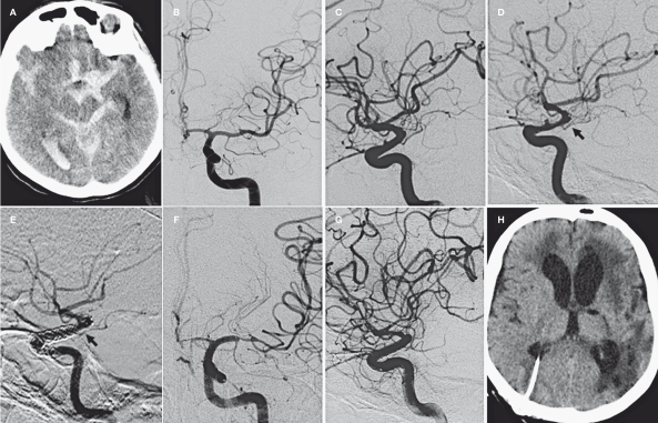
Figure 2.
Case 2. A) CT scan on admission showing subarachnoid and intraventricular bleeding. B-C) Acute AP and lateral view angiogram of the left carotid artery. D) 48 hour pre-treatment angiogram shows a modification of the BBL aneurysm and vasospasm of the carotid siphon and its branches. E) 24 hour control angiogram. Double telescopic Silk stents in the carotid siphon and Solitaire stent in M1 segment. Complete disappearance of the aneurysm. The AChA is still patent (black arrow). F) 7 day control angiogram. Severe vasospasm of ACA and MCA that spared the entire stented arterial segment. G-H) 2 month control angiogram and CT scan. Absence of the AChA and stable occlusion of the aneurysm. Infarct areas in AChA and MCA territories.

