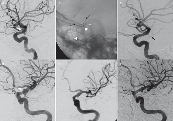
Figure 3.
Case 3. A-B) Different projections of the left carotid siphon showing aneurysm of the C6 segments (white arrows). C) Triple telescopic Silk stent in the carotid siphon. The first stent is placed from M1 to the ophthalmic segment (black arrows), the second one just before the A1-M1 bifurcation (white arrows) and the third one just below the AChA origin (arrowheads). D) Angiographic result at the end of the procedure. E-F) 8 month control angiogram showing a complete disappearance of the BBL aneurysm. Patency of the AChA (black arrow) and narrowing of A1 origin.

