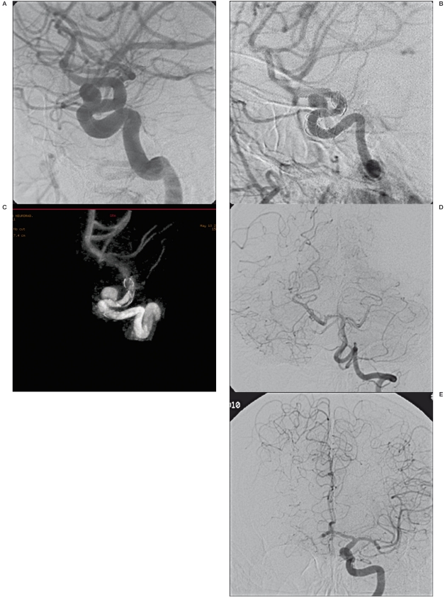
Figure 3.
A-E) Carotid-ophthalmic saccular aneurysm of the left carotid artery (A). Images “b” and “c” show neointimal hypertrophy (arrow) and incorrect stent opening (ring). This condition could be the basis of blood flow changes which prevent aneurysm occlusion. 13 months later, the patient developed a good haemodynamic compensation without clinical symptoms.

