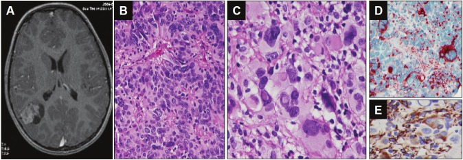
Figure 1.
Neuroimaging and histological findings of the tumor from case 1. A. Axial gadolinium-enhanced T1-weighted MRI image demonstrated a heterogeneously enhanced solid and cystic mass in right parietal lobe. B. Perivascular pseudorosette (Hematoxylin and eosin 200X). C. The giant cell with eosinophilic cytoplasm, eccentrically located single or multiple nuclei with prominent nucleoli, and intranuclear cytoplasmic inclusions (Hematoxylin and eosin, 400X). D. EMA immunohistochemical stain showed perinuclear dot-like or ring pattern. E. GFAP immunohistochemical stain showed positivity of tumor cells.

