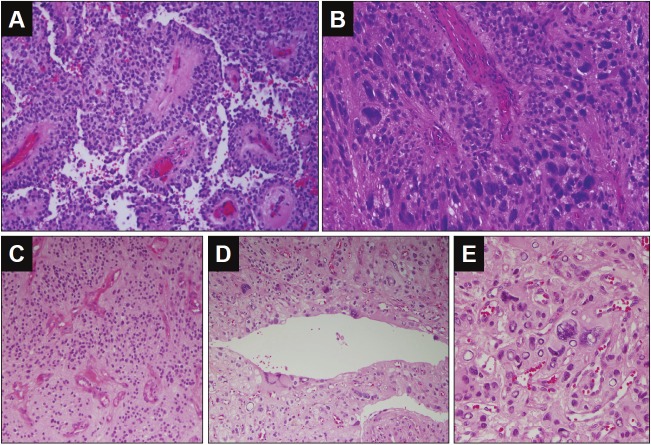
Figure 2.
Histological findings of the tumor from case 2 and case 3. A-B for case 2: A. Low power view of perivascular pseudorosette and pseudo-papillary structure (Hematoxylin and eosin 200X). B. Perivascular pseudorosette formed with pleomorphic tumor cells (Hematoxylin and eosin, 400X). C-E for case 3: C. Low power view of perivascular pseudorosette in well-differentiated area of this tumor (Hematoxylin and eosin 100X). D. Ependymal epithelial surfaces formed with pleomorphic tumor cells (Hematoxylin and eosin, 200X). E. Pleomorphic giant tumor cells. (Hematoxylin and eosin, 400X).

