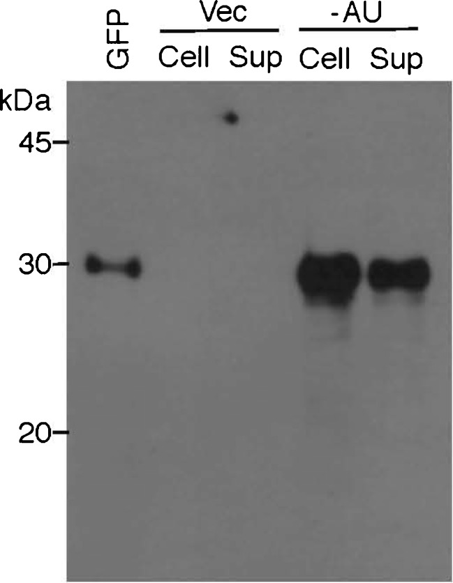
Fig. 11.
Western blot analysis for confirmation of overexpression and easy recovery of GFP from autolysed ABRG5-3 transconjugant cells. The ABRG5-3 transconjugants harboring pVZ321 (Vec) or pGFP461c (−AU) were grown in the CB medium under continuous white light illumination (100 μE m-2 s−1) at 30 °C with shaking (50 rpm) in Erlenmeyer flasks (50 ml of medium in a 300-ml flask exposed to 2 % CO2 gas) for 7 days. After this phase of cell growth for overproduction of GFP, the cell culture (50 ml) was poured into a screw-cap tube and allowed to stand for 3 days on a laboratory bench at room temperature for promoting auto-cell lysis. Cell sediment (100 μl) corresponding to the 50-ml cell culture or protein precipitate (50 μl) from the supernatant corresponding to 10 ml of the 50-ml culture (see details described in “Materials and methods”) was mixed with an equal volume of ×2 SDS sample buffer, then subjected to 12.5 % SDS-PAGE. The gel was also subjected to Western blot analysis with the GFP antibody as in Fig. 4. Purified GFP (29 kDa) protein (1 μg) was also applied as a size marker (left lane)

