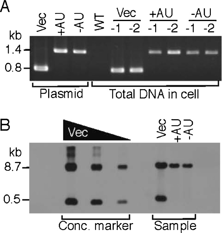
Fig. 2.
Analysis of PCC 6803 transconjugants containing GFP expression vectors. a PCR. Total DNA (1 μg) was isolated from the PCC6803 wild-type (WT) or recombinant cells conjugated with pVZ321 (Vec), pGFP500 (+AU), and pGFP4561c (−AU), respectively. Each two samples (−1 and −2) were subjected to PCR with a set of specific primers, VZ-F2 and VZ-R (Fig. 1). The 1.4- or 0.8-kb position for the PCR-amplified fragments from the pGFP500/461c or pVZ321 plasmid DNA is shown as a positive control at the left. b Southern blot analysis. Total DNA (20 μg) was isolated from the cells in (a), digested by the restriction enzymes HindIII and XhoI, and subjected to Southern hybridization with a specific probe (812 bp, a PCR-fragment amplified with pVZ321 and the primers VZ-F2 and VZ-R). The 8.7- or 0.5-kb position for the concentration marker is indicated at the left. The DNA of pVZ321 (left lane, 0.24 μg, 0.040 pmol; middle lane, 0.080 μg, 0.0133 pmol; right lane, 0.02 μg, 0.0033 pmol) digested with the same restriction enzymes was subjected to hybridization for the concentration marker which was used for a trial calculation of plasmid copy numbers in the cells (Table 2)

