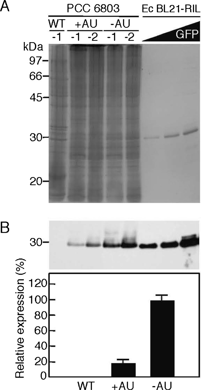
Fig. 4.
GFP expression at the protein level in PCC6803. a SDS-PAGE. Total cellular protein (40 μg) was prepared from wild-type (WT) Synechocystis sp. PCC6803 cells or cells harboring pGFP500 (+AU) or pGFP461c (−AU) grown in the CB medium for 12 days, then each two samples (−1 and −2) were subjected to 12.5 % SDS-PAGE. The gel was stained with Coomassie Brilliant Blue R-250. Purified GFP (29 kDa) proteins (left lane, 1.45 μg, 50 pmol; middle lane, 2.90 μg, 100 pmol; right lane, 8.70 μg, 300 pmol) obtained from E. coli BL21 (RIL as codon plus) harboring pGLO were also applied as concentration markers. The positions (in kilodaltons) from a standard molecular size marker are shown at the left. b Western blot analysis. Top Aliquots of total cellular protein (40 μg) loaded from the samples shown in the different lanes in Fig. 4a in the same order were subjected to Western blotting with a specific antibody for GFP. The 30-kDa position is indicated at the left. Signal intensities corresponding to GFP on an X-ray film in the top panel were measured and presented as relative values (pVZ461c as 100 %) with error bars (n = 3, means ± SD)

