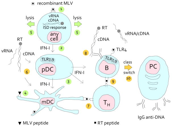
FIGURE 1.
Activation of an anti-DNA specific B cell by mutated endogenous retrovirus. NZB and NZW mice produce endogenous, infectious, replication competent, but xenotropic MLV (76), which cannot infect mouse cells unless pseudotyped with ecotropic or polytropic envelope. These mice also encode many MLVs that are defective in various other ways. In addition, NZW mice also produce ecotropic MLV (77, 78), although it is inefficiently activated from their cells. In the B/W mice, this diversity can lead to phenotypic mixing and generate recombinant viruses. We suggest that some recombinant viruses provide peptides that bind to MHC class I or class II and that are recognized as non-self. As a consequence, B cells will be killed, helped, or both (depending on whether or not they are infected with recombinant virus), resulting in lymphopenia, hyperproliferation and/or autoimmunity. MLV may also infect most other cells, including myeloid dendritic cells (mDC) and plasmacytoid dendritic cells (pDC).
Because the T cells are tolerant to the endogenous virus early in life, sufficiently different recombinant virus has to be generated before cells are killed. In the scheme, the steps leading to lysis are numbered 1 to 5 and colored green. In step 1, recombinant MLV (circle with filled core) enters a hematopoietic or non-hematopoietic cell. If the cDNA fails to integrate, it is stabilized by circularization and triggers the type I interferon stimulatory DNA (ISD) response. (Containing double-stranded RNA, the tRNA-viral RNA (vRNA) priming complex may also be sensed by TLR3; and In B lymphocytes, the vRNA is sensed by TLR7). As a result, type I interferon (IFN-I) is secreted (step 2), which stimulates the plasmacytoid dendritic cell (pDC) to secrete copious amounts of IFN-I, which, in turn, stimulates mDCs (step 3). The mDC presents MLV peptides (step 4) to T helper cells (not shown) that help cytotoxic T cells (not shown) to lyse the MLV-infected cell (step 5).
With the lysis of the infected cell, retroviral cDNA complexed with (mutated) reverse transcriptase (RT) is released (step 6; this and the following steps depict the events that lead to anti-DNA antibody formation and are colored yellow). The cDNA complexes are taken up by pDCs, which secrete IFN to help B and T cells, and by mDCs, which license T helper (TH) cells (step 7). T cell help for B cells with DNA specificity is available due to a recombination event that generates a class II-binding epitope of the RT (RT peptide) that is sufficiently different from self. In step 8, a B cell receptor specific to DNA endocytoses cDNA complexed with RT and presents an RT peptide on MHC class II (step 9). (In a variation on the theme, instead of cDNA, a retro-viral protein mimetope of DNA is bound to the B cell receptor.) This activates the B cell, and after class switching (step 10) the plasma cell produces antibodies to dsDNA. The cognate immune response is helped by the innate immune response to (mutated or non-mutated) retroviral envelope via TLR4 (48, 79); to ss retroviral RNA (vRNA) via TLR7 (53), sensed as part of an endocytosed virus; and to cDNA, via the ISD response (60) or TLR9.

