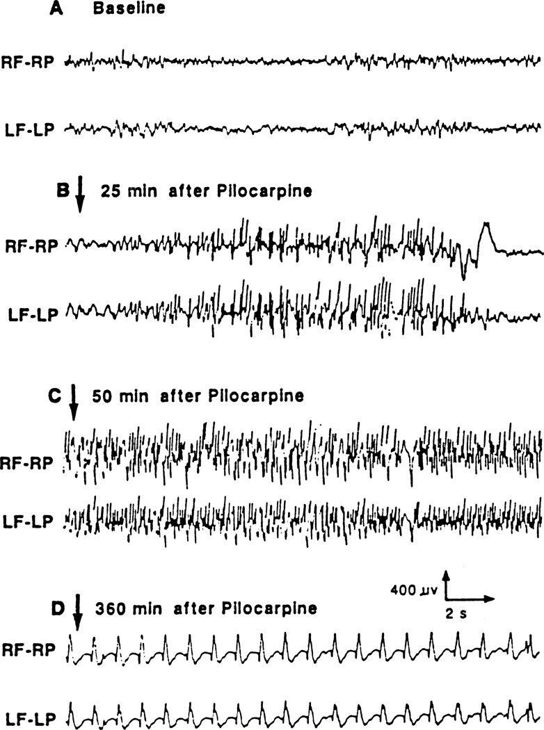
Fig. 2.
Typical example of cortical EEG recording of seizure activity induced by the administration of lithium (3 meq/kg) and pilocarpine (60 mg/kg) in the adult rat. The EEG was recorded through frontoparietal epidural electrodes. Abbreviations: RF–RP: right frontal–right parietal, LF–LP: left frontal–left parietal.

