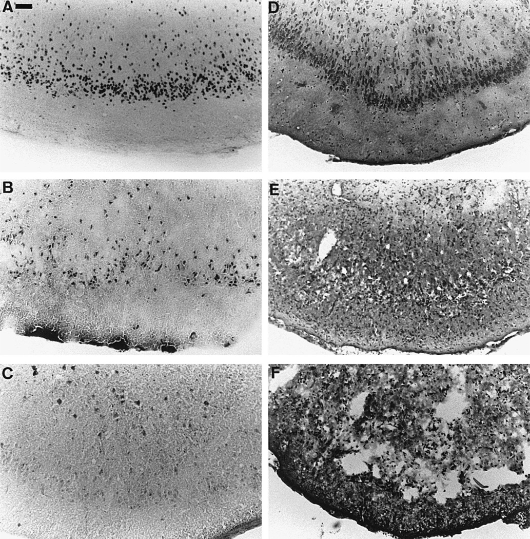
Fig. 5.
Expression of activation, stress and injury markers in sections taken at the level of the piriform cortex. (A) expression of Fos protein in animals sacrificed after 4 h of SE, (B) acid fuchsin staining in animals subjected to 8 h of SE and sacrificed 24 h after SE onset, (C) HSP72 immunoreactivity in animals exposed to 8 h of SE and sacrificed 24 h after SE onset, (D) Cresyl violet staining in control rats, (E) Cresyl violet staining in animals subjected to 8 h of SE and sacrificed 24 h after SE onset and (F) Cresyl violet staining in rats undergoing 8 h SE and sacrificed 144 h after SE onset. Note the high number of nuclei expressing Fos in layer IV of the piriform cortex (A), together with a quite elevated number of neurons stained with acid fuchsin in that area (B) where neuronal damage is also highest (E and F). Conversely, HSP72 is not expressed in layer IV of the piriform cortex, and there are only a few scattered HSP72 positive nuclei in the deeper layers of the cortex (C). Note also the extensive neuronal damage with a complete disorganization and destruction of the piriform cortex in the animals subjected to complete SE and sacrificed 144 h later (F) compared to more limited damage in those sacrificed at 24 h (E). Scale bar: 60 µm.

