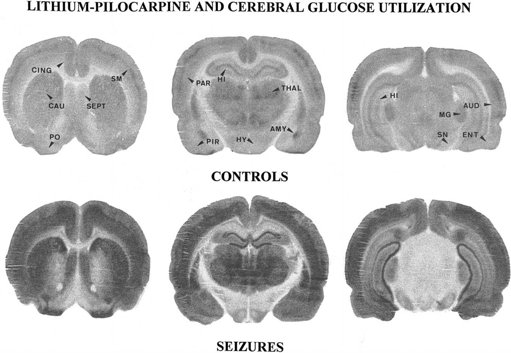
Fig. 7.
Autoradiographic sections taken at the level of the caudate nucleus (CAU), the hippocampus (HI) and the substantia nigra (SN) of control and lithium–pilocarpine exposed rats. Compared to control animals, the grain density was largely increased especially in the whole neocortex, the piriform (PIR) and the primary olfactory cortex (PO), as well as in caudate nucleus, amygdala (AMY), thalamus (THAL), hippocampus and septum (SEPT). In the dorsal hippocampus, the increase was prominent in dentate gyrus (DG), while the pyramidal cell layer of the CA3 area was no longer visible. A large increase in tracer levels also occurred in thalamus (THAL) and substantia nigra, while grain density increased moderately in ventromedian hypothalamus (HY) and medial geniculate body (MG). Abbreviations: CING: anterior cingulate cortex, SM: sensorimotor cortex, PAR: parietal cortex, AUD: auditory cortex, ENT: entorhinal cortex.

