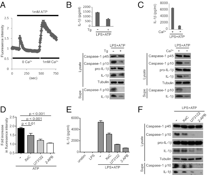
Fig. 1.
Extracellular ATP mobilizes Ca2+ to activate the NLRP3 inflammasome. (A) LPS-primed BMDMs were loaded with Fura-2 followed by stimulation with 1 mM ATP and analysis of Ca2+ flux by time-lapse microscopy. Ca2+ is released from intracellular stores during the initial stimulation in Ca2+-free buffer, and exchange to 1 mM Ca2+-containing buffer permits extracellular Ca2+ influx. (B and C) LPS-primed BMDMs were treated or not with Tg for 30 min to deplete ER Ca2+ (B) or switched to Ca2+ free (−) or Ca2+-containing (+) media immediately before ATP stimulation (C). NLRP3 inflammasome activation was assessed by Western blotting of lysates and supernatants (supes) as well as IL-1β ELISA. (D–F) BMDMs were pretreated with XeC, U73122, or 2-APB, followed by ATP stimulation and analysis of Ca2+ flux (D) and NLRP3 inflammasome activation (E and F).

