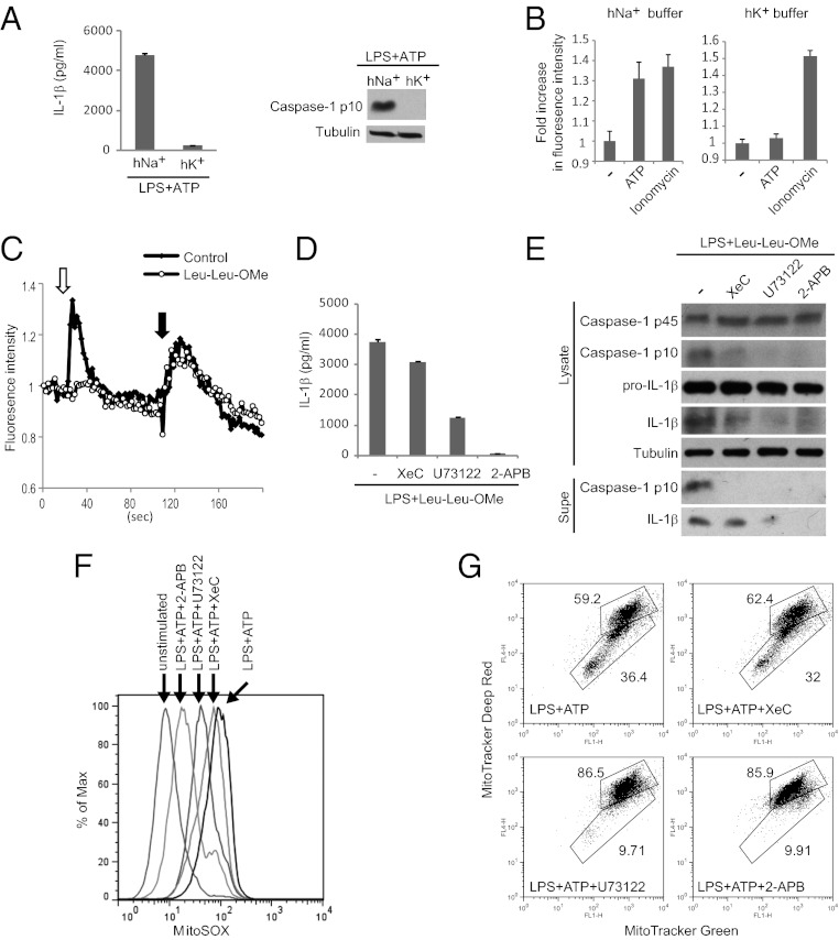
Fig. 3.
Ca2+ signaling promotes mitochondrial damage during ATP stimulation. (A) BMDMs were stimulated with ATP in the presence of 40 mM K+ (hK+) or Na+ (hNa+), followed by analysis of NLRP3 inflammasome activation. (B) Ca2+ imaging of ATP-stimulated BMDMs in the presence of hK+ or hNa+ buffers. Fold-increase represents the ratio of maximal fluorescence to baseline fluorescence. Ionomycin stimulation was included as a control for Fura-2 loading. (C) BMDMs were pretreated with Leu-Leu-OMe or not (control), followed 1 h later by stimulation with 100 μM ATP in Ca2+-free HBSS (white arrow) to induce ER Ca2+ release through the P2Y receptor. Stimulation with the Ca2+ ionophore Ionomycin (black arrow) mobilized Ca2+ from distinct Ca2+ stores resistant to Leu-Leu-OMe and P2YR activation and served as a positive control for Fura-2 loading. (D and E) BMDMs, stimulated as indicated, were examined for NLRP3 inflammasome activation. (F and G) BMDMs stimulated as indicated were stained with MitoSOX (F) or Mitotracker Green and Deep Red (G) followed by flow cytometry analysis.

