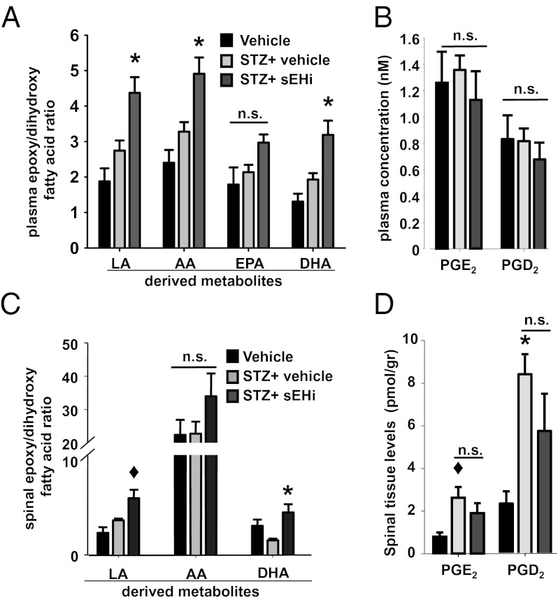
Fig. 3.
Inhibition of sEH modulates levels of EpFAs and dihydroxy fatty acids in plasma and spinal cord tissue. (A) Type I diabetes does not change the plasma levels of substrates and products of sEH (pre-STZ n = 6, post-STZ n = 18 and post-STZ+TPAU n = 6, one-way ANOVA, P = 0.1 for LA and AA metabolites, P = 0.07 for EPA metabolites and P = 0.09 for DHA metabolites). TPAU significantly alters the mean ratio of summed epoxy to dihydroxy fatty acids in diabetic rats, with the exception of EPA metabolites (Kruskal–Wallis one-way ANOVA followed by Dunn’s all pair-wise comparison, *P < 0.02). Table S1 displays the identity and detected quantity of the metabolites analyzed from plasma. (B) Plasma levels of two major prostanoids show no significant change between groups (one-way ANOVA, P = 0.62). (C) Inhibition of sEH leads to selective changes in the levels of spinal EpFAs and their degradation products, dihydroxy fatty acids expressed as ratios (pre-STZ n = 3, post-STZ n = 4, and post-STZ+TUPS n = 6, one-way ANOVA followed by Student Newman–Keuls test; ◆, P = 0.03 for post-STZ vs. post-STZ+TUPS for LA metabolites and *, P = 0.02 for DHA metabolites). (D) Spinal prostanoids are elevated by diabetes but not reduced by inhibition of sEH (one-way ANOVA followed by Student Newman–Keuls test, ◆, P = 0.03 for vehicle vs. post-STZ PGE2 and *, P = 0.006 for vehicle vs. post-STZ PGD2).

