
Fig. 2.
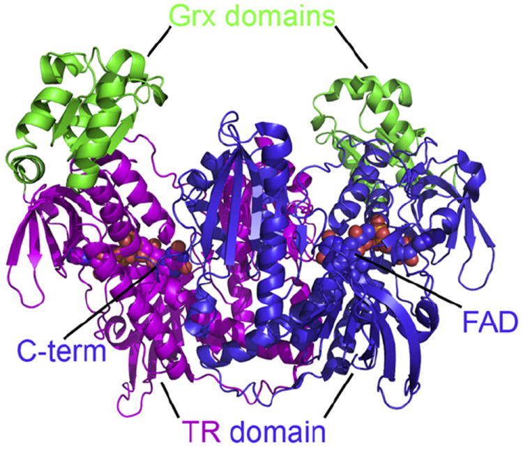
Overall structural architecture of the biological unit of TGR from Schistosoma mansoni. The enzyme is a W-shaped homodimer, each monomer being the fusion of a Grx domain (in green) and a TrxR domain (in magenta and in blue). At the bottom of a large cavity lies the FAD cofactor (shown as a sphere). The C-terminal, Sec-containing arm of each TrxR subunit points towards the FAD binding site of the other.