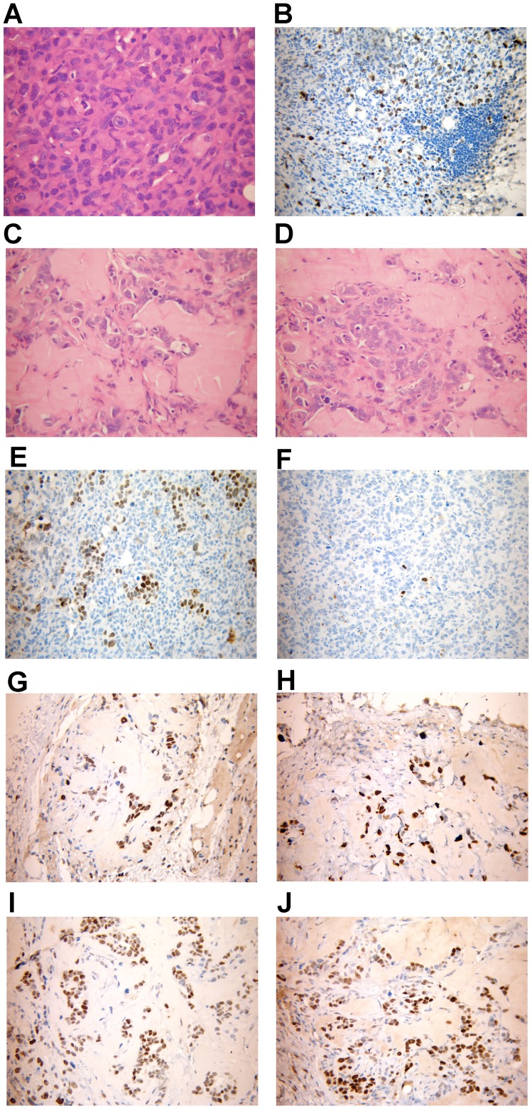
Figure 3. Histopathology and Immunohistochemical analysis of MCF-7 Tet-Off/ACSL4 tumor xenografts.
Histological analysis of human breast tumors formed 70 days after injection of 5×10⁶ MCF-7 Tet-Off/ACSL4 cells into the right flank of female Balb/c nu/nu mice, aged 6–8 weeks. Panel A, C and D showed a representative hematoxylin & eosin stained tissue sections of MCF-7 Tet-Off/ACSL4, MCF-7 Tet-Off empty vector, and MCF-7 Tet-Off/ACSL4 plus doxycyclin treated tumors respectively. Panel B showed a representative immunohistochemical analysis of Ki-67 stained of MCF-7 Tet-Off/ACSL4 tumors. Tumor specimens were stained for detection of ERα and PR receptor expression using the specific antibodies as described in Materials and Methods. Panels showed a representative inmunohistochemical analysis of ERα and PR of the tumor from MCF-7 Tet-Off/ACSL4 xenografts (panel 3E and 3F respectively); from MCF-7 Tet-Off empty vector (panel 3G and 3H respectively) and from MCF-7 Tet-Off/ACSL4 xenografts treted with doxycycline (panel 3I and 3J respectively.

