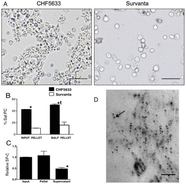
Figure 8. Increased Uptake of CHF5633 by Alveolar Monocyte.
(A) Cell pellet from BALF isolated by 10 min centrifugation at 284× g. The microphotographs were without fixation or staining. Extremely large and soft aggregate of CHF5633 was recovered in the pellet together with cells. Detected Survanta in the cell pellet was minimum after short and low-speed centrifugation and only the cells were detected. Scale bar: 50 µm. (B) Sat PC in pellet and supernatant of input surfactants and BALFs were analyzed. Over 40% of CHF5633 were large and heavy aggregates and were recovered in the pellet. *p<0.001 vs. Survanta. The percent Sat PC in the pellet from the CHF5633 group BALF was higher than that of Input CHF5633. tp<0.05 vs. input CHF5633 pellet. Some of the standard error bars are within the mean value bars. (C) SP-C in pellet and supernatant samples of BALF from CHF5633 group, containing 6.5nmol Sat PC were analyzed by Western blot. SP-C in CHF5633 input sample was given a value of 1. SP-C relative to Sat PC was decreased in supernatant or smaller form surfactant to half of that in input sample and BALF pellet samples. *p<0.05 vs. others. (D) In the surfactant pellet of BALF from the CHF5633 group, a large number of immunogold-labeled SP-C particles (arrow) were associated with surfactant lipid vesicles. Image is representative of findings in n = 3 lambs. Scale bar: 0.1 µm.

