Figure 2.
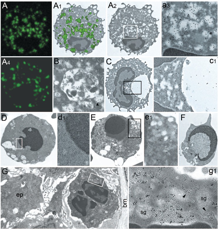
Correlative confocal and electron microscopy of PaCS in SDS neutrophils. (A, 3,000x) Confocal microscopy of an SDS neutrophil immunostained with FK2 antibody against ubiquitinated proteins. Note intensely immunofluorescent areas which, when projected on a TEM micrograph at the same enlargement (A1, 3,000x), show substantial overlapping with PaCS (A2, 3,000x) as confirmed by the intense FK1 immunogold reactivity at higher enlargement (a3, 20,000x). (A4, 3,000x) 20S proteasome immunofluorescence of cytoplasmic areas in another neutrophil from the same SDS patient. (B and C) Autophagic vacuoles and PaCS in SDS neutrophils. Note in B (10,000x) the FK1 reactivity of PaCS, one of which (arrowhead) is apparently discharging its content into the vacuole, and in C (3,000x; enlarged in c1, 20,000x), the p62 reactivity of the vacuole and cytoplasm with, however, no reactivity of PaCS. (D–F, 3,000x) Three apoptotic neutrophils with typical chromatin condensation and segregation (target-type in E and crescentic in F) from SDS patients with SBDS gene mutations. In D, enlarged in d1 (10,000x), cytoplasmic homogenization with disappearance of organelles structure can be seen, whereas in E, enlarged in e1 (10,000x), secretory granules and some FK1 immunoreactivity are still recognizable. Note cytoplasmic vacuoles (of autophagic origin?) in all apoptotic cells. (G, 2,500x) Intraepithelial neutrophil in a gastric biopsy specimen from a patient with H. pylori gastritis, enlarged in g1 (36,000x) to show several PaCS intensely reactive for H. pylori urease. ep: epithelial cell; bm: basal membrane; sg: secretory granules; arrowheads: ribosomes.

