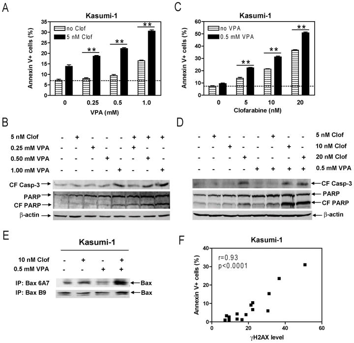
Figure 4. Cooperative induction of apoptosis by clofarabine and VPA in Kasumi-1 cells.
Panels A–D: Kasumi-1 cells were treated with variable concentrations of VPA and fixed concentration of clofarabine (Panels A&B) or variable concentrations of clofarabine and fixed concentration of VPA (Panels C&D) alone or in combination for 72 h. Early and late apoptotic events in the cells post drug treatments were determined by annexin V/PI staining and flow cytometry analysis (Panels A&C). Whole cell lysates were extracted and subjected to Western blotting probed by anti-cleaved caspase-3 (CF casp-3), -PARP, or -actin antibody (Panels B&D). Panel E: Whole cell lysates from Kasumi-1 cells treated with clofarabine and VPA, alone or combined for 72 h, were subjected to immunoprecipitation with the Bax 6A7 (active Bax) or B9 (total Bax) antibody, as described in the Materials and Methods. The immunoprecipitated proteins were subjected to Western blotting probed by anti-Bax or -actin antibody. Panel F: The relationship between the levels for γH2AX and apoptosis in Kasumi-1 cells treated with clofarabine and VPA, alone or combined, were determined by the Pearson test. CF, cleaved form; Clof, clofarabine; ** indicates p<0.005.

