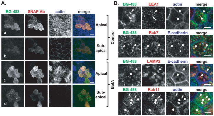
Figure 5. Crumbs3a accumulates intracellularly in the presence of inhibition of the endosomal-lysosomal pathway.
A) Confocal immunofluorescence images of SNAP-Crb3a cells grown on transwell filters, labeled in culture with BG-488, treated for 8 hours with either 100 nM bafilomycin A1 (BafA, panels c and d) or vehicle control (panels a and b), then fixed. Cells were immunostained (not permeabilized) with anti-SNAP antibody and phalloidin. Z-stack images captured at the apical and sub-apical regions (relative to actin-laden microvilli) are shown. White bars, 10 μm. B) Confocal immunofluorescence images of cells treated with bafilomycin A1 as in 5A, but cells were permeabilized and immunostained with indicated antibodies. In the top three rows, arrows highlight regions of co-localization of internalized, labeled SNAP-Crb3a and antibody of interest. In the bottom row, heavy arrows (pointing to right) highlight internalized, labeled SNAP-Crb3a from the cell surface while the light arrows (pointing to left) highlight Rab11-positive vesicles. White bars, 5 μm.

