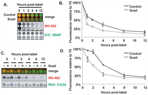
Figure 7. Snail destabilizes Crumbs3a at the cell surface.
SNAP-Crb3a cells co-expressing Snail or vector control were cell surface labeled with BG-682 and either fixed (A) or lysed (B) at the indicated time points. A) in situ immunocytochemistry showing half-life of cell surface SNAP-Crb3a relative to cell surface SNAP-Crb3a as described in 3A(c), in the presence of Snail-FLAG (Snail) or vector control. B) Quantitation of fluorescence images from (A) showing relative residual fluorescence signal at the specified time points, normalized to the total anti-SNAP antibody signal in each well. C) Immunoblot of cell lysates labeled in parallel to those in 7A with D) quantification of residual fluorescence signal of cell surface SNAP-Crb3a normalized to total cell SNAP-Crb3a performed as described in Figure 3A(d). Experiments were performed twice in duplicate; graphs show averaged experimental time points with standard deviations.

