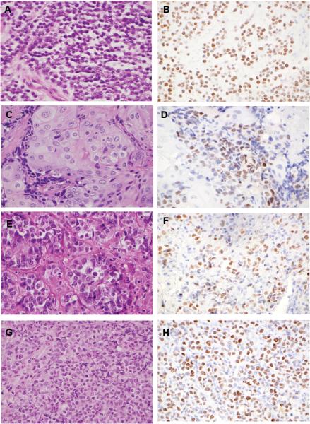
Figure 1. Histopathology and NUT immunohistochemistry.

Representative histologic images and immunohistochemical staining patterns for all 4 newly diagnosed mediastinal NMC cases. Case numbers correspond to the clinicopathologic features outlined in Tables 3 and 4: panels A and B, Case 1; panels C and D, Case 2; panels E and F, Case 3; panels G and H, Case 4.
