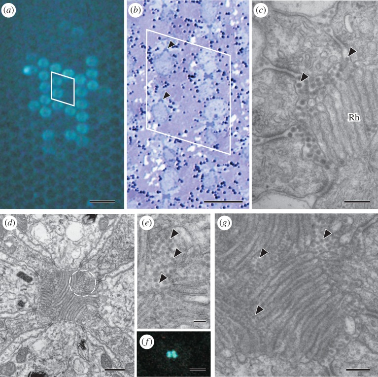
Figure 4.
Fluorescing ommatidia in the eye of P. glacialis. (a) UV epi-illumination reveals fluorescence in a subset of ommatidia. (b) A semi-thin, light-microscopical section of the eye observed in (a). The arrowheads indicate the same two fluorescing ommatidia in (a). The unequally sized cell bodies of R1 and R2 show that the ommatidia are of type I; in this particular case, type Ia. (c) The extracellular space of the rhabdom (Rh) of type I is filled with particles of diameter 30–40 nm. (d) A type II ommatidium of Papilio xuthus. Broken circle indicates one of four clusters of particles. (e) High magnification picture of the circled region in (d). Arrowheads indicate particles. (f) A transverse view of a section of an ommatidium with four fluorescing dots in the centre. (g) Part of a rhabdom of a fluorescing ommatidium of G. sarpedon. Particles are found within the rhabdom area. Scale bars: (a) 50 µm, (b) 20 µm, (c) 0.2 µm, (d) 0.50 µm, (e) 0.1 µm, (f) 5 µm, and (g) 0.2 µm.

