
FIG. 5.
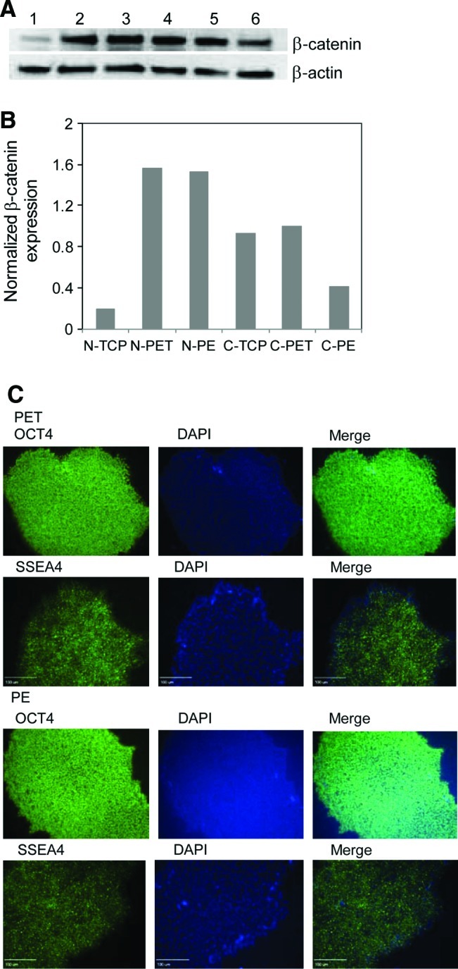
Translocation of β-catenin to nucleus of H9 cells grown on membrane substrates. (A) Western blotting analysis of β-catenin expression in nucleus (lanes 1–3) and cytoplasm (lanes 4–6) of hESCs grown on TCP substrates (lanes 1 and 4), PET (lanes 2 and 5), and PE (lanes 3 and 6) membrane substrates. (B) Semiquantification of β-catenin in nucleus (N) and cytoplasm (C) through western blotting assay. The Kodak 1D gel imaging analysis software was used to perform these semiquantification analyses. β-Actin served as a control for semiquantification. (C) Detection of pluripotency of hESCs after culturing cells on PET and PE membranes for 2 days. Cells were stained with antibodies against OCT4 and SSEA4 marker proteins and also labeled with DAPI. The labeled cells were examined under a fluorescence microscope (Olympus IX 71) equipped with a CCD camera. Scar bar: 100 μm. Color images available online at www.liebertonline.com/tea