
Figure 1.
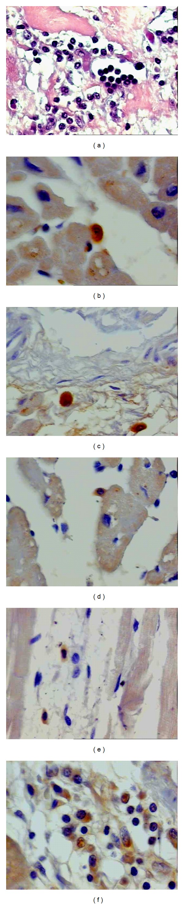
Histological sections of heart tissues obtained at autopsy from subjects with chronic chagasic cardiomyopathy. (a) Cellular exudation with a predominance of mononuclear cells around the fibrosis area (1,280X); (b) positive TNF-α staining and leukocytes in close contact with the myocardiocyte (1,280X); (c) positive IFN-γ staining in the inflammatory exudate (1,280X); (d) positive IL-4 staining in the inflammatory exudate and leukocytes in close contact with the myocardiocyte (600X); (e) discrete TGF-β immunostaining in the inflammatory exudate (1,280X); (f) positive NOS2 staining in the inflammatory exudate (1600X).