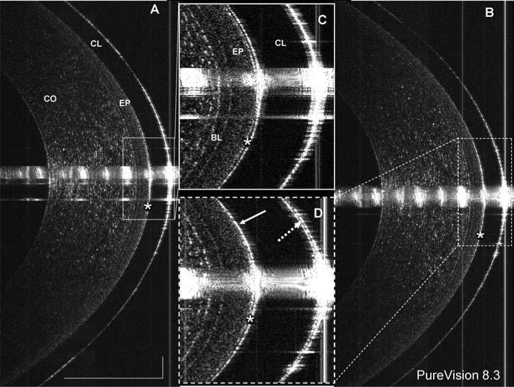
FIG. 1.
Direct visualization of tears on a contact lens. A PureVision lens with base curve of 8.3 mm was fitted onto the eye. Using ultra-high resolution OCT with an 8 mm scan width, images were taken at the center of the cornea (A) immediately after lens insertion and (B) after one blink. The post-lens tear film (*) was clearly visualized in the highly magnified images (C and D). The post-lens tear film was 6 μm thick immediately after lens insertion (C) and 5 μm thick after the blink (D). After the blink (B and D), the pre-lens tear film (dotted arrow) was 5 μm thick. Interestingly, after the blink, the post-lens tear film was not visualized in the upper portion of the cornea (solid arrow in D), possibly due to pressure exerted by the lid during the blink. The bars denote 500 μm for two images (A and B).

