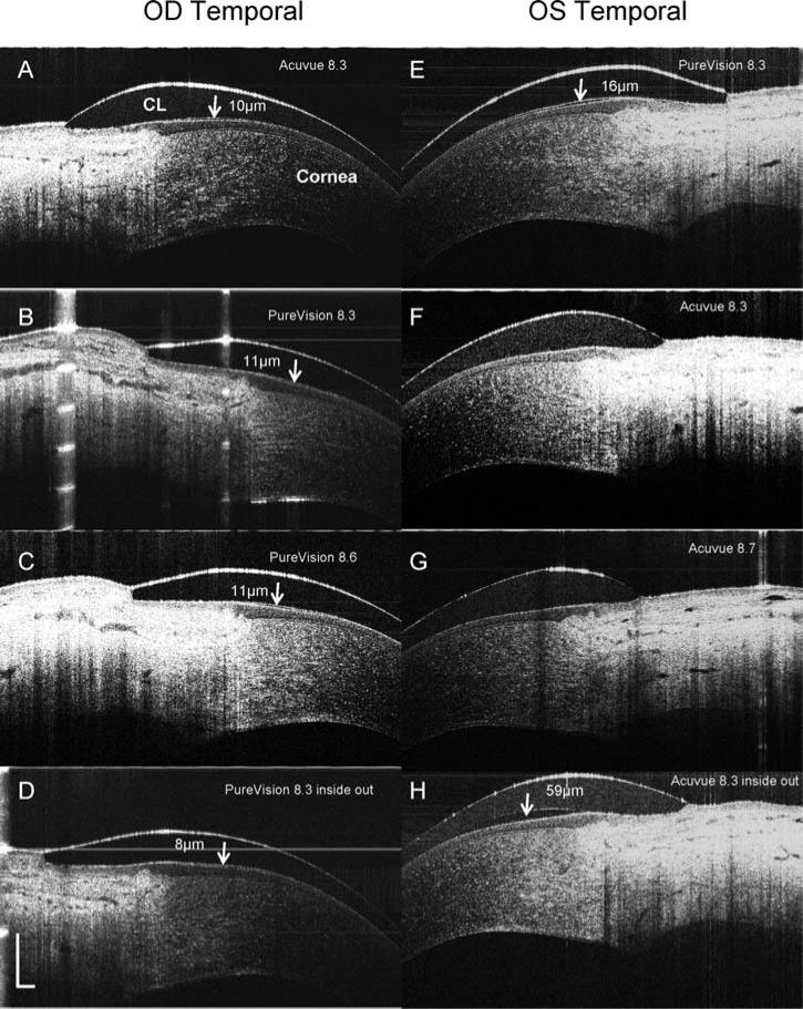
FIG. 2.
OCT images of contact lens edges in situ. PureVision (E, B, C, D) and ACUVUE Advance (A, F, G, H) lenses with different base curves were fitted on the eyes of a subject. The lens edges were visualized in OCT images that were taken from the temporal side when the subject looked at a nasally located target. OCT scanning with an 8 mm scan width was directed at a right angle to the edge of the lens (CL). The post-lens tear film was visualized with the PureVision lens images (B, C, D, and E) and in some ACUVUE lens images (A and H). After the lenses were fitted inside out (D and H), the post-lens tear film was visualized with both. Note the configurations of the lens edges are different with a round edge in the Pure-Vision and a sharp edge in the ACUVUE lens. The thickness of the post-lens tear films were measured at the locations marked (arrows). Bars = 500 μm.

