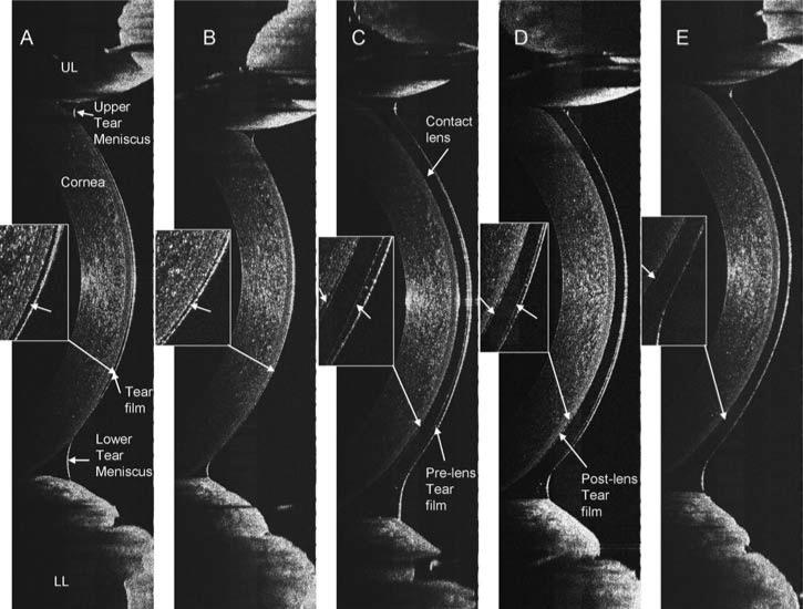
FIG. 6.
The tears on the ocular surface and contact lens. A vertical 12 mm OCT scan was performed. After the instillation of one drop of the artificial tears, the tear film on the cornea without a lens and tear menisci around the upper (UL) and lower (LL) eyelids were clearly visualized (A with enlarged inset), and recovery was evident 5 min after the instillation (B with enlarged inset). On the same eye, a PureVision lens was fitted, and one drop of the artificial tears was instilled. The pre- and post-lens tear films (arrows) were imaged (C–E with enlarged insets). Tear menisci were located around the upper and lower eyelids. Note that the boundary between the tear and lens was not clearly visualized, due to the lower light scattering. Two minutes after the insertion, the tear films were still visualized, especially underneath the lower part of the lens (E).

