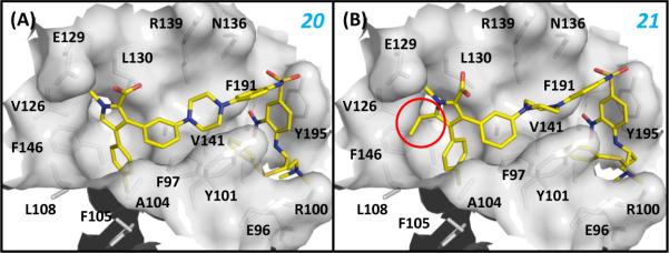
Figure 8.
Binding models between (A) 20, (B) 21 and Bcl-XL. The Bcl-XL protein from the crystal structure between 1 and Bcl-XL was used in the docking simulations. The highest ranked poses of both compounds were selected as the binding models. The added ethyl group to 20 was denoted by the red circle. Residues of Bcl-XL at the binding site were labeled.

