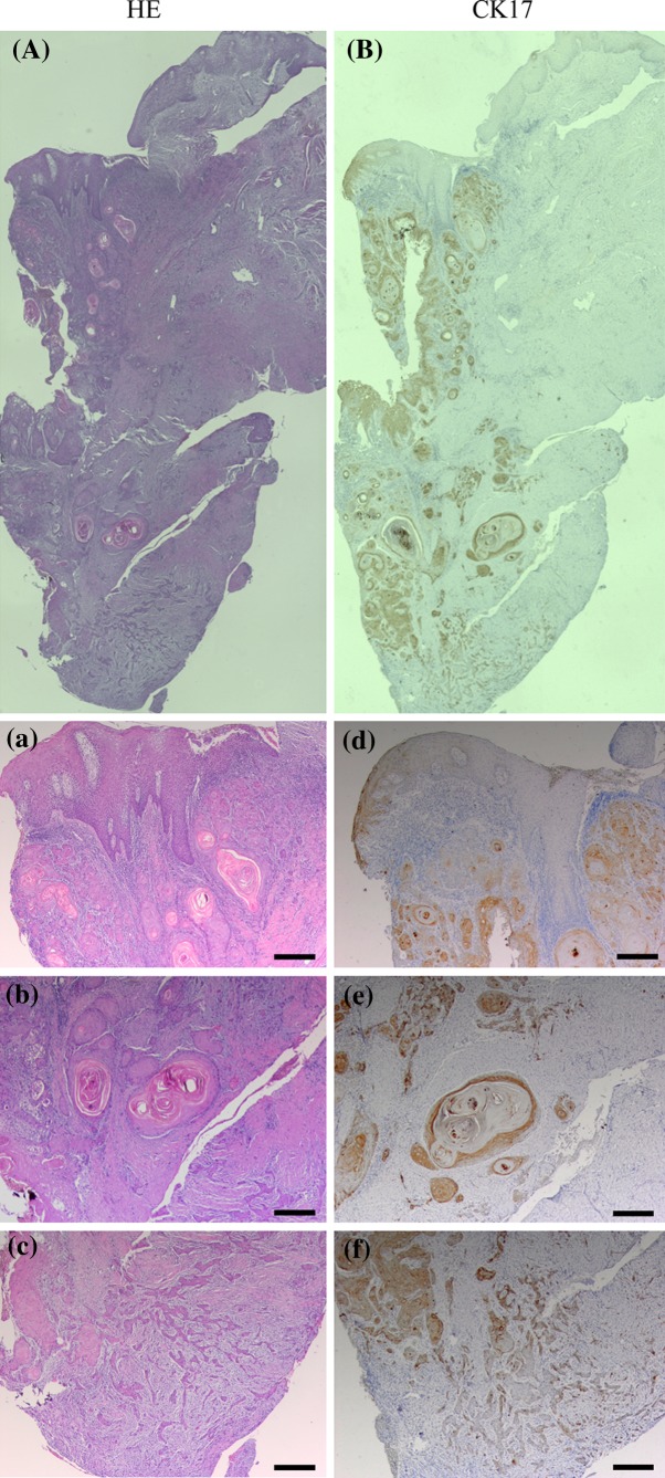
Fig. 2.
A representative case of OSCC with dysplasia, cancer nests, and infiltrating cells. Serial sections were stained with HE (A, a–c) and anti-CK17 antibody (B, d–f). High-magnification views of selected areas are shown (a–f). CK17 was markedly expressed in dysplasia, while not in normal epithelium (d). CK17 was obviously expressed in the inner layer of cancer nest, while not in the outer (e). CK17 was absent in the infiltrating cells (f). (Original magnification ×40, scale bars 200 μm)

