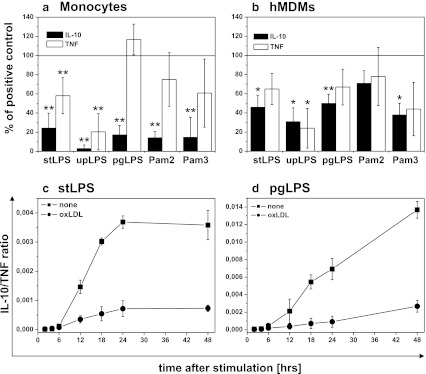
Fig. 2.
Effect of oxidized LDLs on PAMPs-induced IL-10 and TNF production in monocytes and monocyte-derived macrophages (hMDM). a, b Monocytes were isolated from PBMC by adherence and placed in media supplemented with 10 % FCS. hMDMs were differentiated from adherent monocytes for at least 7 days in medium supplemented with 10 % human serum, and before experiment, cells were placed in media supplemented with 10 % FCS. Cells were cultured alone or treated for 30 min with oxidized LDLs at the 15 μg/ml and then stimulated with PAMPs as indicated in the X-axis. Supernatants were collected 20 h after stimulation and IL-10 (black bars) and TNF (white bars) concentrations were determined by ELISA. Data are expressed as percent of positive control that is cells stimulated in the absence of oxLDLs (indicated as 100 %). Values are the mean ± SD from at least five independent experiments. **p < 0.01, *p < 0.05 versus corresponding positive controls. Absolute cytokine levels are presented in supplementary data Table 1. c, d Monocytes were isolated from PBMC by adherence and placed in media supplemented with 10 % FCS. Cells were cultured alone (black squares) or treated for 30 min with oxidized LDLs at the 15 μg/ml (black circles), and then stimulated with stLPS (c) or pgLPS (d). Supernatants were collected at time points indicated in the figure, and IL-10 and TNF concentrations were determined by ELISA. For each time point, IL-10/TNF biological activity ratios recalculated as described in “Materials and Methods” are presented in the figure (c, d). Values are the mean ± SD from three independent experiments. Absolute cytokine levels are presented in supplementary data Figure 1. Similar experiments were done with upLPS, Pam2CSK4, or Pam3CSK4 (data not shown).

