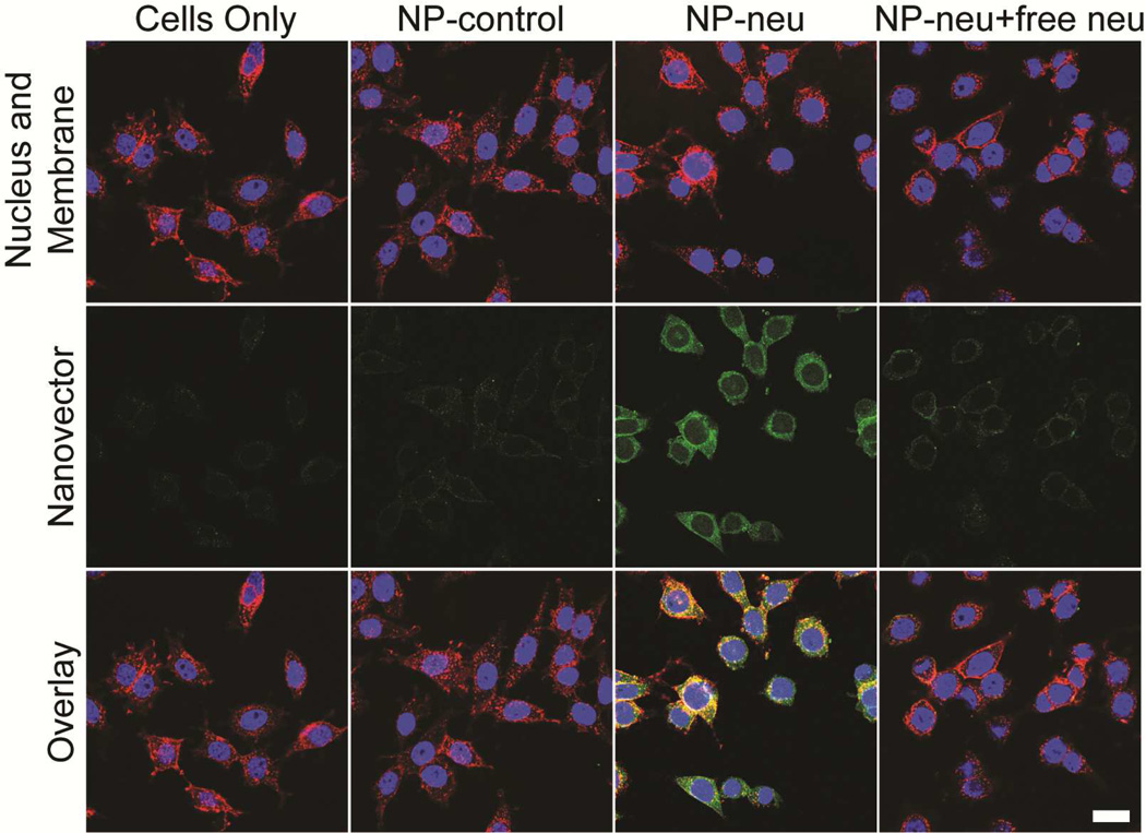
Figure 4.
Confocal imaging of NP treated MMC cells. Cells were treated with non-targeted NP-control, targeted NP-neu, or targeted NP-neu with excess free neu for 2 hrs in fully supplemented culture media. The images of untreated cells (first column) are provided as a reference. Bright green fluorescence is only seen in the NP-neu treated cells indicating these NPs are able to target the neu receptor on neu expressing cells. Image acquisition times were identical for all the samples. Scale bar corresponds to 20 µm.

