
Figure 4.
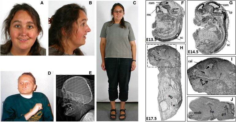
Human Craniofacial Anomalies and Murine Phf21a Expression in Craniofacial and Brain Regions
(A–E) Human craniofacial anomalies with PHF21A truncation and PHF21A deletion.
(A–C) Female subject MCN1762 with balanced translocation t(1;11) exhibits ID and CFAs that include brachycephaly, microcephaly, a long narrow nose, mild midfacial hypoplasia, thin lips, and prominent ear lobes.
(D) The PSS male proband (PSS-Romeike) with a deletion including PHF21A is shown at the age of 31 years. He shows microcephaly, brachycephaly, a broad forehead, a long narrow nose, a hypoplastic mandible, very thin lips, hypotelorism, and dysplastic low set ears, in addition to ID and no speech, among other phenotypes.
(E) A topogram of a cerebral CT of individual PSS-Romeike at the age of 33 years, shortly before he died, shows a hypoplastic mandible and the parietal foramen at the back of the head.
(F–J) Phf21a expression during murine craniofacial development.
(F and G) Expression of Phf21a at embryonic days (E) 13.5 and 14.5. Highest transcript abundance is seen in the developing CNS with [35S]-UTP labeled in situ probes. The following abbreviations are used: rom, roof of midbrain; rnc, roof of neopallial cortex; sc, spinal cord; and cp, intraventricular portion of cerebellar primordiu.
(H and I) At E17.5, prominent signals were detected with Phf21a antisense probes in bones of the facial skeleton. The palatine bone is marked by arrows, and the orbitosphenoidal bone is marked by a black arrowhead. Signals in the intestine were also detected with the sense probes and are most likely not specific (these are marked by asterisks). The following abbreviation is used: cal, calvaria.
(J) Sagittal sections of the adult mouse brain showed expression of Phf21a within the hippocampal formation, the cerebellum, and the olfactory bulb. Three different Phf21a antisense probes gave consistent results. The following abbreviations are used: mob, main olfactory bulb; hp, hippocampus; and gcl, granule cell layer of the cerebellum.