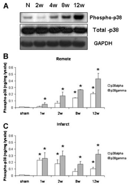
Fig. 1.
A: Representative Western blots indicating phosphorylation (i.e., activation) of p38 MAPK in the remote myocardium during post-MI remodeling. Changes in p38α (white bars) and p38γ (gray bars) were measured via ELISA of tissue lysates from remote (B) or infarct/borderzone myocardium (C). n = 4–9, *P < 0.05, week 8 and 12 versus week 1.

