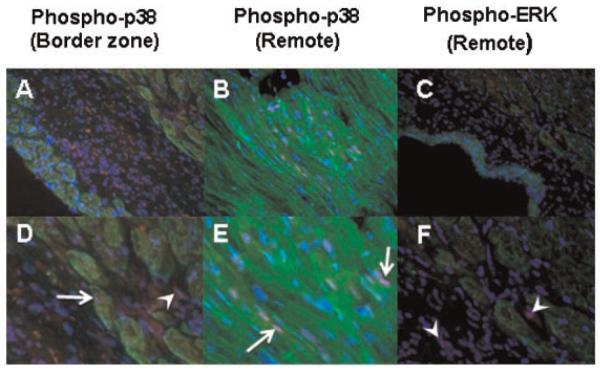
Fig. 4.
Immunofluorescent co-staining of phospho-p38 (red, A,B,D,E) and phospho-ERK (red, C,F) together with myosin heavy chain staining using MF-20 (green). Nuclear staining for phospho-p38 was observed in both myocytes (arrows) and non-myocytes (arrowheads), whereas phospho-ERK staining, even in fibrotic areas of the remote myocardium, predominated in non-myocytes (arrowheads). Nuclei are counterstained with DAPI; cardiac non-myocytes appear black. A–C: 400 ×; D–F: 1,600 ×.

