
Figure 1.
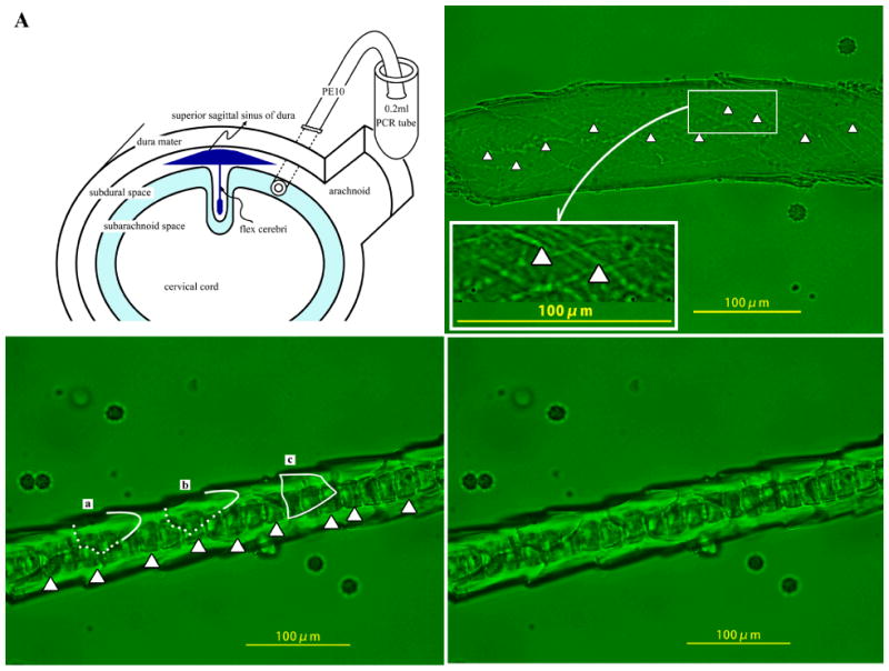
An SP-10 polyethylene tube inserted into rat cisterna magna was used for administration of arachidonic acid and collection of cerebrospinal specimens (A). Capillary of pia mater was observed by fluoromicroscopy 30 min after intracerebrospinal injection of arachidonic acid in rats administered with (C-2) or without (B) RCN9 cells. Arrowheads show the endothelial cell junction (B). Contour of RCN9 cells is traced (C-1).