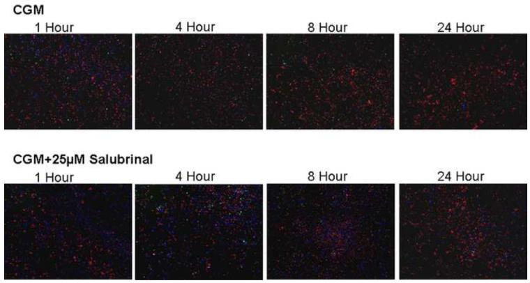
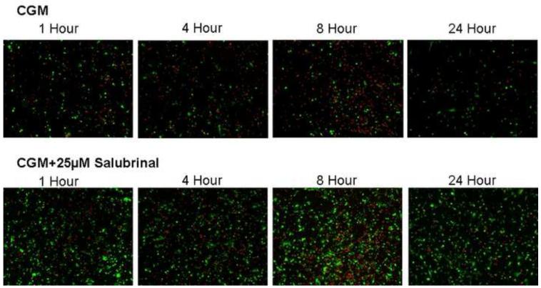
Figure 5. Time Course Fluorescent Microscopy Assessment of UPR Inhibition on HCEC Cells Following Hypothermic Storage.
Fluorescent images were taken 1, 4, 8 and 24 hours post-storage following 24 hours of cold storage in Complete Media (CGM) with either 0μM or 25 μM salubrinal. (A) Blue labeled (Hoechst) cells denote living cells, red (propidium iodide) are necrotic and green (Yo-Pro-1) indicates apoptotic cells. The micrographs corroborated the trends in cell death as determined with flow cytometric analysis with salubrinal addition resulting in increased viable cell and total cell retention populations. (B) Green labeled (calcein-AM) cells indicate viable cells and red (propidium iodide) cells denote necrotic cells. The micrographs demonstrate that salubrinal addition resulted in increased cellular membrane integrity, cell attachment and overall cell viability throughout the initial 24 hours post-expsoure.


