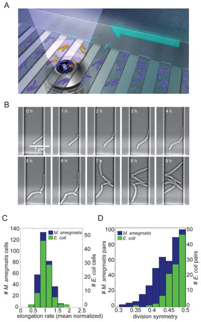
Figure 1. M. smegmatis exhibits heterogeneous growth characteristics.
(A) Schematic diagram of the microfluidic device used for long-term imaging of mycobacteria. Media flows through the main channel (large arrow) and provides nutrients (cyan circles) by diffusion (small arrows) to the cells. (B) Bright-field, time-lapse imaging of M. smegmatis in the microfluidic device.. (C) Distribution of average (mean centered) elongation rates of 322 individual M. smegmatis (blue; left axis) and 102 E. coli (green; right axis) cells averaged over the course of one cell cycle. The mean elongation rates were 1.15 μm/h for M. smegmatis and 3.72 μm/h for E. coli. (D) Distribution of division symmetry for 166 M. smegmatis (blue; left axis) and 105 E. coli (green; right axis) pairs of sister cells. Division symmetry is calculated as the ratio of the length of the smaller sister to the sum of the lengths of both sisters at division.

