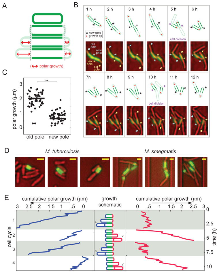
Figure 2. M. smegmatis growth is asymmetric and elongation occurs from the old pole.
(A) Schematic diagram of the pulse-chase experiment used to measure polar growth. Cells were labeled with amine reactive dye (green) and growth was assessed by measuring the extension of the unlabeled region (red arrows). Cell wall-labeling did not alter cell elongation rate (fig. S5A), other labeling chemistries such as hydroxylamine labeling via periodate oxidation led to similar staining patterns (data not shown), and similar labeling patterns were seen in cells grown in broth and in microfluidic channels (fig. S5B). (B) Time-lapse imaging of two sister cells following the pulse-labeling (green) of the cell wall. The bright field images were pseudo-colored red. New poles are annotated with asterisks and the old poles with arrows. Each cell’s poles are annotated with the same color asterisk and arrow. A schematic is drawn above each image, marking the new poles and growth poles with an asterisk and red arrow, respectively. We also used a fluorescein-vancomycin conjugate (Van-FL) to stain nascent peptidoglycan (30) and found preferential labeling of the old pole over the new pole (fig. S5C). (C) Growth over one cell cycle at new versus old poles in 50 cells (*p<0.001 by a Mann-Whitney rank sum test). There was no cell in which the new pole elongated more than the old pole. (D) Three representative images of M. tuberculosis (left) and M. smegmatis (right) following the pulse-labeling (green) of the cell wall (after 48 hours in broth culture for M. tuberculosis and six hours in the microfluidic device for M. smegmatis). The bright field images are pseudo-colored red. Size bars represent 1.3 μm. Comparison images of E. coli labeled under similar conditions may be found in fig. S5D. (E) Cumulative polar growth for one labeled cell plotted for each pole through four cell cycles. The cell inheriting the new pole alternates the direction of growth after division events.

